
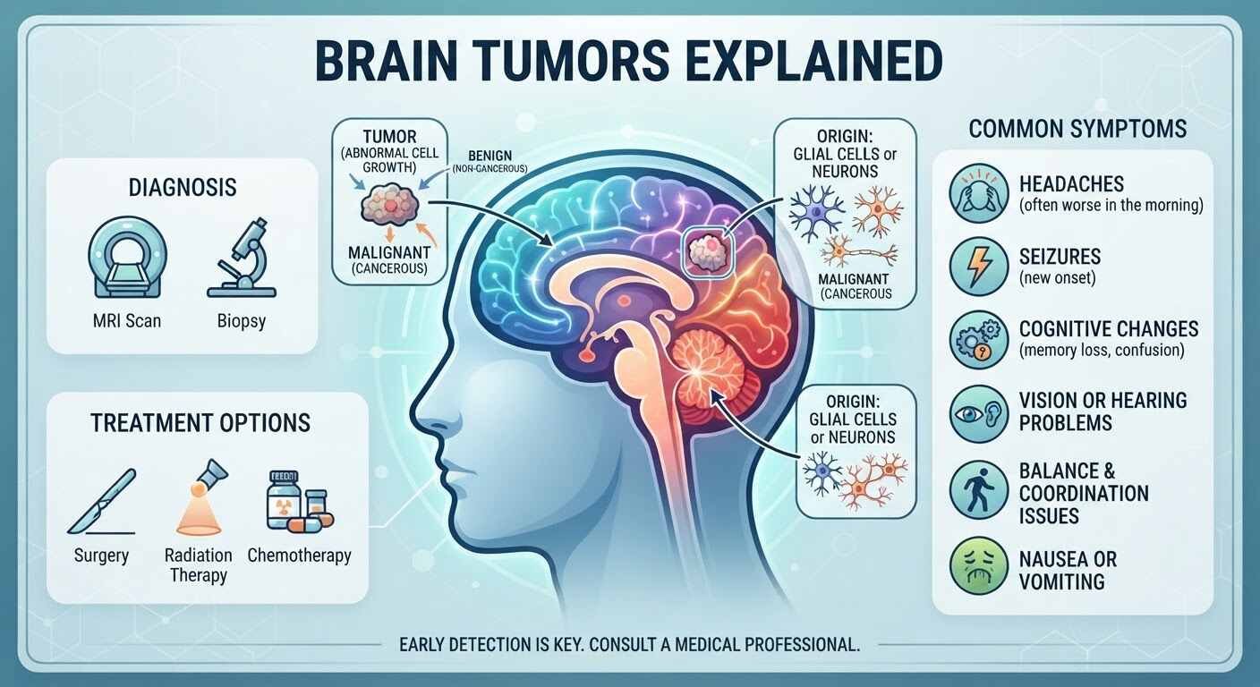
A brain tumor diagnosis can feel overwhelming, whether it involves you or a loved one. The complexity of the human central nervous system means that brain tumors present unique challenges compared to cancers in other parts of the body. Understanding the basics—what brain tumors are, how they are classified, what warning signs to watch for, and how modern medicine treats them—is an essential first step toward navigating a path forward.
What Is a Brain Tumor?
A brain tumor is an abnormal growth or mass of cells within or surrounding the brain. Inside the rigid structure of the skull, any uncontrolled growth can cause significant problems. As a tumor enlarges, it increases pressure inside the cranium (intracranial pressure) and can compress, invade, or damage healthy brain tissue.
When dealing with complex brain or spine conditions, consulting a leading expert like Dr. Simranjeet Singh—widely recognized as a best neurosurgeon in guwahati—ensures that patients receive accurate evaluations and personalized treatment plans.
Primary vs. Secondary Brain Tumors
Brain tumors broadly fall into two main categories based on where they originate:
- Primary Brain Tumors: These originate directly within the brain or its immediate surrounding tissues, such as the meninges (membranes covering the brain), cranial nerves, or pituitary gland. Primary tumors can be benign (non-cancerous) or malignant (cancerous).
- Secondary (Metastatic) Brain Tumors: These occur when cancer elsewhere in the body—most commonly the lungs, breast, colon, kidney, or skin (melanoma)—spreads through the bloodstream to the brain. Metastatic brain tumors are far more common than primary tumors and are always malignant.
Key Types of Primary Brain Tumors
Primary brain tumors are named according to the type of cell from which they develop.
Gliomas
Gliomas account for nearly 30% of all primary brain tumors and 80% of malignant ones. They arise from glial cells, which support and protect neurons. The main sub-types include:
- Astrocytomas: Originating from star-shaped cells called astrocytes. They range from slow-growing (Grade 1) to highly aggressive (Grade 4).
- Glioblastomas (GBM): The most common and aggressive malignant primary brain tumor in adults. Glioblastomas grow rapidly and readily invade surrounding brain tissue.
- Oligodendrogliomas: Arising from oligodendrocytes (cells that insulate nerve fibers), these tend to grow more slowly than astrocytomas.
- Ependymomas: Developing from cells lining the brain’s ventricles and central canal of the spinal cord, often interfering with cerebrospinal fluid (CSF) flow.
Meningiomas
Meningiomas arise from the meninges, the protective membranes wrapping the brain and spinal cord. They represent the most common primary brain tumor in adults. The vast majority (over 80%) are benign and grow slowly, though their size can eventually exert pressure on critical tissue.
Pituitary Adenomas
Located at the base of the brain, the pituitary gland controls hormone production. Adenomas here are almost exclusively benign. However, as they grow, they can disrupt hormone regulation or press against the nearby optic chiasm, causing visual field loss.
Acoustic Neuromas (Schwannomas)
These benign tumors develop on the vestibular nerve, which connects the inner ear to the brain. They cause gradual hearing loss, tinnitus (ringing in the ear), and balance issues.
Understanding Brain Tumor Grading
Unlike cancers elsewhere in the body, which are staged (Stages I–IV) based on how far they have spread, primary brain tumors are assigned a Grade (Grades 1–4) by the World Health Organization (WHO). Grading reflects how aggressive the tumor cells look under a microscope and how quickly the tumor is expected to grow and spread into surrounding tissue.
| WHO Grade | Cell Characteristics | Growth Rate | Common Examples |
| Grade 1 | Benign, slow-growing cells; look almost normal | Very Slow | Pilocytic Astrocytoma, Meningioma |
| Grade 2 | Slightly abnormal cells; can recur or progress | Slow | Low-grade Oligodendroglioma |
| Grade 3 | Malignant, actively reproducing abnormal cells | Moderate to Fast | Anaplastic Astrocytoma |
| Grade 4 | Highly malignant, abnormal cells with rapid blood vessel growth and dead tissue (necrosis) | Rapid & Invasive | Glioblastoma |
Symptoms to Watch For
Because different areas of the brain control different bodily functions, symptoms depend heavily on the tumor’s size, growth rate, and location.
General Symptoms
General symptoms occur due to increased pressure inside the skull:
- Persistent or Worsening Headaches: Particularly headaches that are worse in the morning, wake you from sleep, or worsen with coughing or bending over.
- Seizures: A sudden onset of seizures in an adult with no prior history is a primary warning sign.
- Nausea and Vomiting: Unexplained, often severe in the morning, and unrelated to gastrointestinal issues.
- Cognitive Changes: Confusion, memory lapses, difficulties concentrating, or personality shifts.
Location-Specific Symptoms
- Frontal Lobe: Changes in judgment, emotional regulation, motor impairment on one side of the body, or speech difficulties.
- Temporal Lobe: Memory issues, difficulty understanding language, or sensory hallucinations (smell/taste).
- Parietal Lobe: Sensory disturbances, difficulty with spatial orientation, or trouble recognizing familiar objects.
- Occipital Lobe: Partial or complete loss of vision, visual hallucinations, or field deficits.
- Cerebellum: Unsteadiness, loss of coordination, difficulty with fine motor skills, or slurred speech.
- Brainstem: Facial weakness, double vision, swallowing difficulties, and vital function disruptions.
Diagnostic Methods
When a brain tumor is suspected, a multidisciplinary medical team uses several specialized tests to confirm the diagnosis:
- Neurological Examination: Testing reflexes, muscle strength, balance, coordination, vision, hearing, and cognitive function.
- Magnetic Resonance Imaging (MRI): The gold standard imaging technique. Advanced MRI protocols—such as functional MRI (fMRI), magnetic resonance spectroscopy (MRS), and perfusion MRI—map functional pathways and evaluate tumor blood flow.
- Computed Tomography (CT) Scan: Often used in emergency settings to detect bleeding, calcification, or bone involvement.
- Biopsy: Obtaining a tissue sample (either during surgical removal or via a stereotactic needle biopsy) is essential for definitive molecular diagnosis and grading.
Modern Treatment Modalities
Brain tumor management requires a personalized, multidisciplinary approach involving neurosurgeons, neuro-oncologists, radiation oncologists, and rehabilitation specialists.
1. Surgery
Surgery is frequently the first line of treatment when a tumor is safely accessible.
- Goals: Achieve maximum safe resection (removing as much tumor as possible without damaging crucial brain function) or perform a biopsy.
- Techniques: Modern neurosurgery utilizes image-guided navigation, intraoperative MRI, awake craniotomies (for tumors near speech or motor centers), and fluorescence-guided surgery to maximize removal while protecting healthy tissue.
Consulting with an experienced practitioner like Dr. Simranjeet Singh—renowned as a best neurosurgeon in guwahati—provides access to world-class neurosurgical care, precise diagnostic evaluations, and individualized treatment strategies designed for the best possible quality of life.
2. Radiation Therapy
High-energy rays are used to destroy tumor cells or stop them from growing.
- External Beam Radiation (EBRT): Targeted radiation delivered over several weeks.
- Stereotactic Radiosurgery (SRS): Techniques like Gamma Knife or CyberKnife deliver a single, highly precise dose of radiation to a concentrated area, minimizing damage to surrounding tissue.
3. Chemotherapy
Chemotherapy uses systemic drugs to kill rapidly dividing cells.
- A primary challenge in treating brain tumors is overcoming the blood-brain barrier—a protective network of blood vessels that restricts substances from entering brain tissue.
- Temozolomide (TMZ) is an oral chemotherapy drug that successfully crosses this barrier and is widely used for high-grade gliomas.
4. Targeted and Tumor-Treating Fields (TTFields)
- Targeted Therapy: Focuses on specific genetic mutations or pathways within cancer cells (e.g., BRAF inhibitors or anti-angiogenic agents like Bevacizumab that block blood supply to tumors).
- Tumor-Treating Fields (Optune): A non-invasive device worn on the scalp that creates low-intensity electric fields to disrupt cell division in glioblastoma cells.
Looking Ahead
Advances in neuro-oncology are accelerating rapidly. Genomic profiling and molecular markers (such as IDH mutation status and MGMT promoter methylation) now allow doctors to tailor treatments precisely to an individual tumor’s profile. Emerging therapies, including personalized cancer vaccines and immunotherapies, offer promising new avenues for managing complex diagnoses.
If you or someone you care about experiences new, persistent neurological symptoms, prompt medical evaluation is vital. Early diagnosis remains key to expanding treatment options and improving outcomes.
When facing complex brain or spine conditions, patient outcomes heavily depend on early diagnosis and precision surgical techniques. Seeking guidance from a trusted specialist—such as finding a best neuro doctor in guwahati —ensures access to state-of-the-art diagnostic protocols and surgical safety. Experienced specialists like Dr. Simranjeet Singh utilize modern micro-neurosurgical tools and intraoperative monitoring to protect healthy brain function during surgery.