
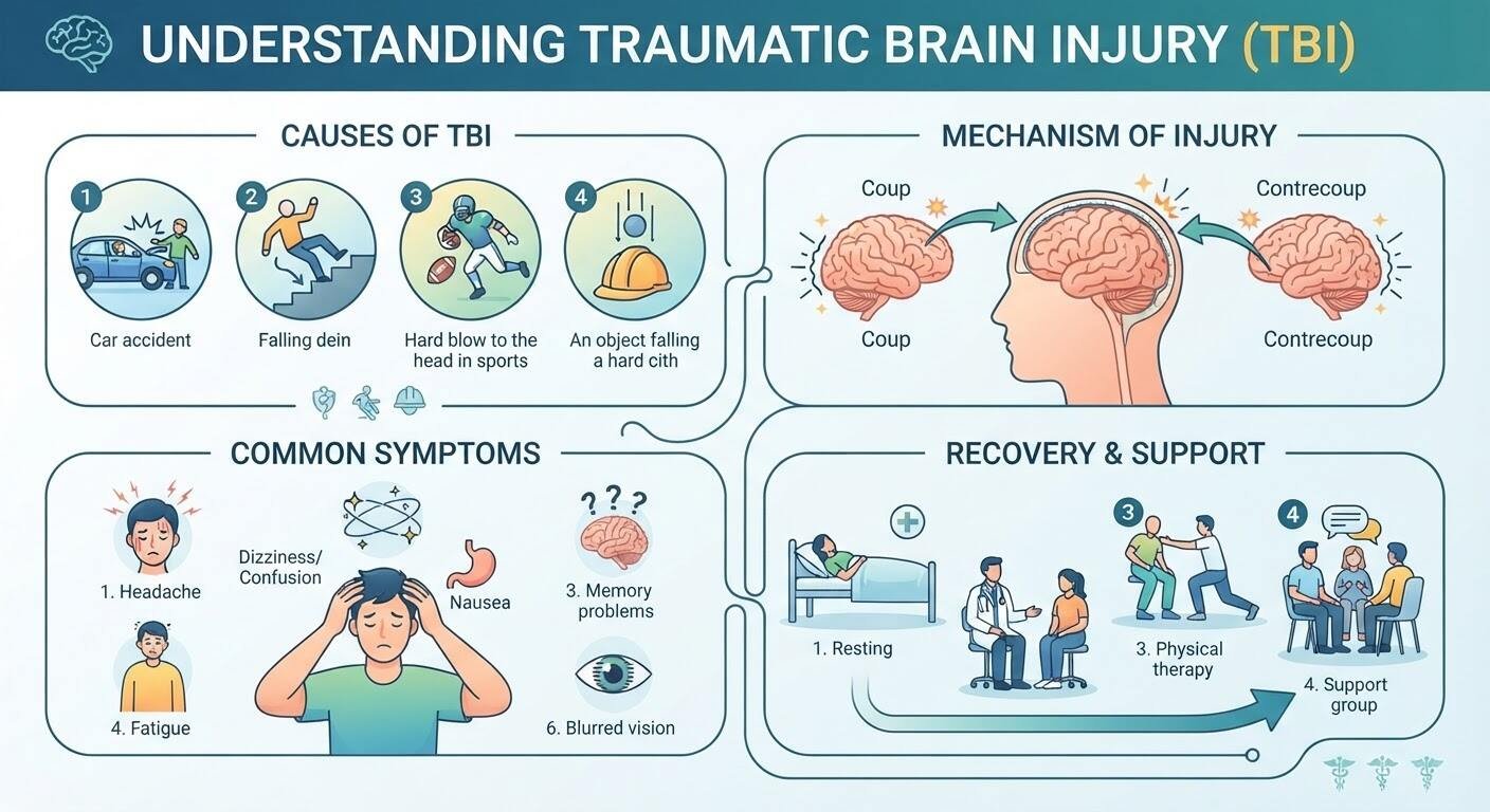
A blow to the head is one of the most common injuries in daily life. It can happen in a split second during a road accident, an unexpected slip on wet stairs, a workplace incident, or a rough collision on a sports field. Because our skull is hard and made of solid bone, we often believe that if there is no outer cut or bleeding, the brain inside is completely safe.
However, inside the skull, the human brain has a soft, delicate texture similar to firm gelatin. It rests inside a bath of protective fluid called cerebrospinal fluid. When a sudden and violent impact hits the head, the brain shifts rapidly, bouncing or twisting against the rough inner ridges of the bony skull. This abrupt movement can stretch brain tissue, bruise delicate structures, tear fragile blood vessels, and disrupt nerve pathways that control how we move, talk, and think.
In medicine, this injury is known as a Traumatic Brain Injury (TBI). A TBI is not a single, simple wound. It is a spectrum of injuries ranging from a mild concussion to deep, life-threatening brain bleeding. Because early symptoms often feel like an ordinary headache or brief tiredness, people frequently ignore them until serious complications develop.
Knowing what happens during a head injury, watching for hidden warning signs, and consulting an experienced clinical specialist—such as seeking an evaluation with the Best Neurosurgeon in Guwahati—is the safest and most effective way to protect your long-term health and brain function.
What Really Happens to the Brain During an Impact?
To understand how a brain injury develops, it helps to understand how damage unfolds in two distinct stages: primary injury and secondary injury.
The Primary Injury: The Instant of Contact
The primary injury occurs at the precise millisecond of physical contact. This is the direct mechanical damage caused by the hit. Depending on the direction and speed of the blow, the damage may include:
- Direct bruising of the outer cortex of the brain.
- Tearing of microscopic blood vessels, causing blood to escape into tissue.
- Shearing or stretching of delicate nerve fibers (axons) across different lobes.
- Direct bone displacement if a skull fracture drives pieces of bone downward.
The Secondary Injury: The Chain Reaction
The damage does not always stop once the accident ends. Over the following hours, days, and even weeks, a chain reaction of chemical and physical changes can occur inside the head.
Injured brain cells release stress chemicals, triggering rapid inflammation and swelling (cerebral edema). Because the adult skull is an unyielding bone box that cannot stretch or expand, swelling brain tissue has nowhere to go.
As swelling increases, intracranial pressure (ICP)—the pressure inside the skull—rises dangerously. High pressure squeezes blood vessels flat, cutting off fresh oxygen and glucose supplies to healthy brain tissue. Without urgent surgical or medical steps to lower this pressure, healthy brain cells begin to starve and die.
Common Causes of Traumatic Brain Injuries
Traumatic brain injuries cut across every age group and lifestyle. Understanding the common situations that lead to head trauma helps in prevention and rapid response:
- Road Traffic Collisions: Vehicle accidents involving cars, two-wheelers, and pedestrians account for a massive share of severe head trauma. Rapid stops cause the head to snap back and forth violently.
- Accidental Falls: Slips in bathrooms, falling from ladders, tripping down staircases, or simple balance loss in older adults are leading causes of head trauma and brain bleeds.
- Sports and Recreational Activities: High-impact sports like football, cricket, cycling, and martial arts carry regular risks of concussions and contusions from direct collisions.
- Workplace & Industrial Mishaps: Construction sites and industrial plants carry risks of falling objects, high drops, and machinery collisions.
- Physical Assaults and Blunt Force: Direct hits to the skull from domestic assaults, street altercations, or blunt instruments create direct focal brain contusions.
The Main Types and Severity Levels of TBI
Doctors categorize traumatic brain injuries into mild, moderate, and severe categories based on how the patient responds, their level of consciousness, and structural scan results.
1. Concussion (Mild TBI)
A concussion is the most common form of brain injury. Even though it is labeled “mild,” it is a real neurological event. In a concussion, the brain experiences a brief metabolic disruption. Standard CT scans often look completely normal because the injury exists at a chemical and cellular level rather than as a visible large tear.
Patients often feel dazed, dizzy, foggy, or nauseous. Most people recover completely with proper mental and physical rest, but repeated concussions can cause lasting damage.
2. Brain Contusion
A contusion is an actual bruise on the surface of the brain, complete with small broken blood vessels and localized swelling. Contusions often happen directly under the site of impact.
They can also happen on the exact opposite side of the brain. When the skull stops abruptly, the brain rebounds and slams against the far wall of the skull. This two-sided injury is known as a coup-contrecoup injury.
3. Diffuse Axonal Injury (DAI)
Diffuse Axonal Injury is one of the most serious forms of brain trauma. It does not occur in just one isolated spot. Instead, severe rotational forces—such as when a car spins or rolls during an accident—twist and stretch the brain inside the head.
This twisting tears microscopic communication fibers (axons) across wide areas of brain tissue. Because the brain’s internal communication network is cut, DAI is one of the primary reasons patients slip into a coma after an accident.
4. Intracranial Hematomas (Brain Bleeds)
When blood vessels rupture inside the skull, blood escapes and pools into a clot called a hematoma. This trapped blood takes up valuable space, pushing the brain to one side and compressing vital control centers. The three common types include:
- Epidural Hematoma: Bleeding forms between the skull bone and the dura mater (the tough outer membrane protecting the brain), often from a torn artery linked to a skull fracture. Patients frequently experience a misleading “lucid interval”—they are knocked out, wake up feeling fine for a short while, and then rapidly slip into unconsciousness as arterial blood pools quickly.
- Subdural Hematoma: Bleeding occurs underneath the dura mater, usually caused by torn veins. These can be acute (bleeding fast after a hard blow) or chronic (slow, silent oozing over weeks, common in senior citizens after minor falls).
- Intracerebral Hemorrhage: Bleeding occurs directly inside the soft brain tissue itself, damaging local functional networks.
Early Warning Signs You Should Never Overlook
One of the biggest hazards with head trauma is assuming everything is fine just because the person can stand up and speak immediately after the incident. Symptoms often develop gradually over hours as blood pools or tissues swell.
To help you distinguish between mild symptoms that need rest and dangerous signs that demand emergency care, keep this comparison in mind:
| Symptom Group | Early / Mild Indications | Red-Flag Emergency Indicators |
| Headache & Head Sensations | Dull headache, mild tension, fuzzy head feel | Severe, worsening pain; feeling like the head might burst |
| Stomach & Digestive Issues | Mild nausea, lack of appetite | Repeated, forceful vomiting (projectile vomiting) |
| Alertness & Consciousness | Brief daze, tiredness, wanting to rest quietly | Inability to stay awake, fainting, falling into deep coma |
| Vision & Sensory Changes | Mild light sensitivity, slight eye strain | Blurred vision, double vision, unequal pupil sizes |
| Motor & Movement Control | Mild clumsiness, slight unsteadiness | Weakness in an arm or leg, limping, facial drooping |
| Speech & Thinking | Forgetfulness about the hit, mild slowness | Slurred speech, total confusion, inability to form words |
The Critical “Red Flag” Symptoms
If you or anyone around you exhibits any of the following signs after a blow to the head, go to an emergency trauma center immediately:
- One eye’s black pupil is noticeably larger than the other pupil (anisocoria).
- Repeated vomiting that does not stop.
- Clear fluid or watery blood leaking out of the nose or ears (a warning sign of a skull base break leaking brain fluid).
- New-onset seizures, body twitching, or sudden muscle fits.
- Sudden numbness, paralysis, or loss of control on one side of the body.
- Growing confusion, strange aggressive behavior, or slurred, garbled speech.
When red-flag signs show up, every minute counts. Having the patient evaluated by an expert neurotrauma team under the direction of the top neurosurgeon in Guwahati ensures that life-saving surgical steps are taken without dangerous delays.
How Neurosurgeons Diagnose Traumatic Brain Injury
When an injured person arrives at a hospital, the clinical team works through an organized diagnostic pathway to evaluate brain health quickly and safely:
1. Clinical Examination & The Glasgow Coma Scale (GCS)
Doctors check vital signs, assess pupil responses with a light, and calculate a Glasgow Coma Scale (GCS) score. The GCS evaluates three key behaviors:
- Eye Opening: Does the patient open their eyes naturally, only to sound, only to touch, or not at all?
- Verbal Response: Are they oriented to time and place, confused, using random words, or silent?
- Motor Response: Can they follow simple hand commands, pull away from pressure, or show abnormal stiff reflexes?
Scores range from 3 (deep unconsciousness) to 15 (fully awake and responsive).
2. Emergency Non-Contrast CT Scan
The Non-Contrast Computed Tomography (CT) scan is the gold standard for emergency trauma triage. Within minutes, a CT scan provides clear cross-sectional images of the skull bones and brain. It reveals acute blood collections, identifies skull fractures, and shows whether swelling is shifting the brain sideways across its centerline.
3. Detailed Magnetic Resonance Imaging (MRI)
An MRI scan takes longer than a CT, so it is rarely used for unstable emergency patients. However, once the patient is stable, an MRI gives detailed soft-tissue images. Specialized sequences pinpoint microscopic nerve tears (DAI), tiny brainstem contusions, and hidden tissue damage that CT scans cannot catch.
4. Continuous Brain Pressure (ICP) Monitoring
For comatose or severely injured patients in intensive care, neurosurgeons place a tiny pressure probe through a small opening in the skull into the fluid spaces or brain tissue. This monitor provides continuous, real-time pressure readouts, alerting doctors to internal pressure spikes long before visible body symptoms emerge.
Modern Treatment Pathways: Medical Care to Advanced Surgery
Treatment for a traumatic brain injury depends on its severity, location, and the speed at which symptoms evolve. The primary goal is always the same: keep the brain oxygenated, stop active bleeding, control swelling, and prevent secondary tissue damage.
Medical Management & Neuro-Intensive Care
For mild concussions or stable, minor contusions that do not require immediate operations, structured medical care is the primary approach:
- Hyperosmolar Therapy: Doctors give specialized intravenous medicines, such as mannitol or concentrated hypertonic saline. These solutions draw excess water out of swollen brain cells into the bloodstream, lowering pressure inside the skull.
- Controlled Ventilation and Sedation: Patients with severe injuries may be placed on a ventilator. Carefully controlling carbon dioxide and oxygen levels helps regulate cerebral blood vessel diameter, while medical sedation lowers the brain’s metabolic work.
- Anti-Seizure Medications: Irritated, bruised brain tissue can generate electrical misfires. Doctors often prescribe short courses of preventive anti-seizure medicines to stop fits that could otherwise deprive the brain of oxygen.
- Temperature and Glucose Control: Preventing high fevers and stabilizing blood sugar prevents additional cellular stress in healing brain tissue.
Surgical Neuro-Trauma Care
When blood clots push against vital brain regions or internal swelling spikes to dangerous levels, surgical intervention is critical:
- Craniotomy for Clot Evacuation: A neurosurgeon creates a small window in the skull by removing a section of bone (bone flap). The surgeon opens the thick protective membranes, removes the trapped blood clot (hematoma), seals the bleeding blood vessel, and returns the bone flap to its natural place with small titanium plates and screws.
- Decompressive Craniectomy: In cases of massive, widespread brain swelling, the swelling itself can crush the brainstem against the base of the skull. In this life-saving procedure, the neurosurgeon removes a large portion of the skull bone and does not replace it immediately. Instead, the bone is preserved in a medical tissue bank or under the skin of the abdomen. This gives the swelling brain room to expand outward safely beyond the skull walls until the swelling subsides weeks later.
- External Ventricular Drain (EVD): If blood or swelling blocks normal cerebrospinal fluid flow, doctors insert a tiny, soft tube into the fluid ventricles of the brain to drain excess fluid into an outside collection bag, lowering pressure.
- Depressed Skull Fracture Repair: If fractured bone fragments are driven inward toward brain tissue, surgeons lift the bone pieces, clean away debris, repair damaged dura membranes, and rebuild the skull contour.
Rehabilitation: Helping the Brain Rebuild Its Pathways
Surviving the acute phase of a brain injury is the first step; regaining physical, mental, and emotional independence is the journey that follows. The brain has an innate healing ability known as neuroplasticity—the capacity of healthy neurons to learn new tasks and take over functions lost from damaged areas.
A structured rehabilitation program includes:
- Physical Therapy: Focuses on restoring gross motor skills, leg strength, core balance, walking stability, and hand-eye coordination.
- Occupational Therapy: Helps patients relearn practical day-to-day activities such as buttoning clothes, using utensils, preparing meals, typing, and handling basic self-care.
- Speech and Cognitive Therapy: Assists patients who have difficulty finding words, slurring speech, or swallowing food. Therapists also use targeted exercises to strengthen working memory, attention span, and planning skills.
- Neuropsychological and Emotional Support: Frontal lobe trauma frequently leads to unexpected emotional changes—such as irritability, mood swings, anxiety, depression, and low frustration tolerance. Psychological support and family counseling provide coping strategies for these changes.
Preventive Steps for Everyday Safety
While accidents happen without warning, simple everyday habits significantly lower the risk of severe brain trauma:
- Always Wear an Approved Helmet: Whether riding a motorcycle, scooter, or bicycle, wearing a well-fitted helmet absorbs impact forces and reduces traumatic brain injury risk by over 70%.
- Buckle Your Seatbelt Every Trip: Seatbelts keep your body secured, preventing your head from hitting steering wheels, dashboards, or windshields during sudden stops.
- Fall-Proof Living Spaces for Seniors: Install sturdy grab bars in bathrooms, remove loose throw rugs, keep hallways well-lit, and place non-slip mats in showers to prevent severe domestic falls.
- Use Protective Gear in Sports: Children, teenagers, and adult athletes should always use appropriate headgear and helmets during contact sports, skating, and cycling.
- Never Drive Under the Influence: Alcohol and sedating medications slow reaction times, blur depth perception, and remain leading factors in severe vehicular trauma.
Protect Your Brain Health with Prompt Expert Care
The human brain defines our memories, our personality, and everything that makes us who we are. Even what seems like an ordinary bump on the head can hide slowly leaking blood vessels or escalating swelling beneath the skull.
Never ignore progressive headaches, morning vomiting, sudden dizziness, or slight confusion after an impact. Early diagnostic scans and expert clinical oversight provide peace of mind and protect against permanent nerve damage.
If you, an elderly parent, or a loved one have experienced a recent blow to the head and are dealing with lingering or unusual symptoms, seek an immediate, thorough clinical evaluation from the best neurosurgeon in Guwahati, Dr. Sumanjeet Singh. Timely medical attention and skilled neurotrauma care ensure that you or your loved ones receive an accurate diagnosis, focused treatment, and the strongest foundation for a full recovery.